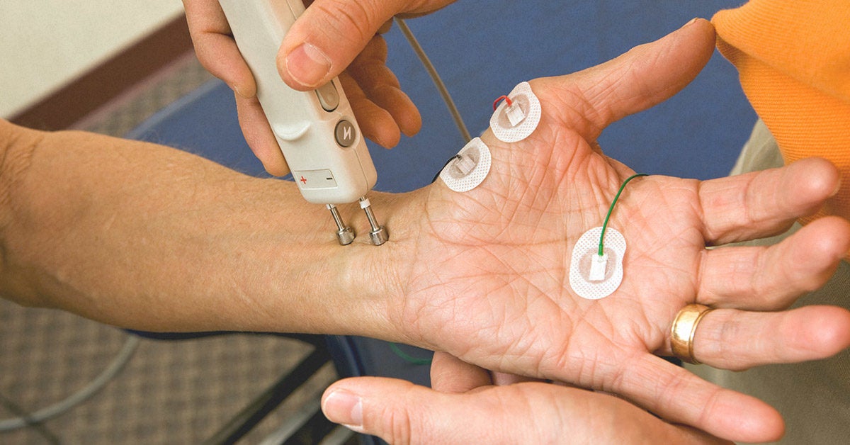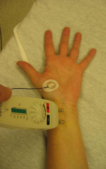

In some cases, cigarettes and caffeinated drinks, such as coffee, tea, and cola may be restricted 2 to 3 hours before testing. Generally, fasting is not needed before the test. Read the form carefully and ask questions if something is not clear. You will be asked to sign a consent form that gives your permission to do the test. Your healthcare provider will explain the test to you, and you can ask questions. Below is a list of common steps that you may be asked to do: Both tests help find diseases that damage the nerves and muscles.Īsk your healthcare provider to tell you what you should do before your test. It's often done during the same visit as the EMG. NCV can determine if your nerves are damaged. NCV measures the speed of conduction of an electrical impulse through a nerve. It's also called a nerve conduction study (NCS). Pinched nerve, such as carpal tunnel syndrome or a pinched nerve in the spine (radiculopathy)Ī related test that is often done is a nerve conduction velocity (NCV).

This is a genetic or immune disorder that occurs at the point where the nerve connects with the muscle.ĭamage or disease of the motor nerve, such as can be seen with nerve disease This is a chronic genetic disease that slowly impairs how muscles work. This is an inflammatory muscle disease that causes decreased muscle power.

When it contracts, its electrical activity may make abnormal patterns.Īn abnormal EMG result may be a sign of a muscle or nerve disorder, such as: But if your muscle is damaged or has lost input from nerves, it may have abnormal electrical activity during rest. As you contract your muscle more forcefully, more and more muscle fibers are activated, making more action potentials.Ī healthy muscle will show no electrical activity (no signs of action potential) during rest. The action potential (size and shape of the wave) that this makes on the monitor gives information about how your muscle responds to nerve stimulation. But after that, there should be no signal.Īfter the electrode is put in, you may be asked to contract your muscle, such as by lifting or bending your leg. When an electrode is put in, a brief period of activity can be seen. Muscle tissue does not normally make electrical signals during rest. The number and location of muscles depends on what the healthcare provider is looking for.ĮMG measures the electrical activity of your muscle during rest, slight contraction, and forceful contraction.

Often, several muscles are tested 1 at a time. An audio-amplifier is used so the activity can be heard. The electrical activity picked up by the electrodes is then displayed on a monitor in the form of waves. The test is used to help find nerve and muscle problems.ĭuring the test, your healthcare provider will put small needles (also called electrodes) through your skin into a muscle. Electromyography (EMG) measures muscle response or electrical activity in response to a nerve’s stimulation of your muscle.


 0 kommentar(er)
